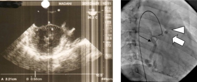
Figure 2.

left: Transesophageal echocardiography shows dilated mitral valve annulus and prolapsed mitral valve leaflets about 6 mm. right: RCA (black arrow) injection at right anterior oblique (RAO) view shows opacification of left coronary artery LCA) (white arrow) and pulmonary artery (arrow head) consecutively.
