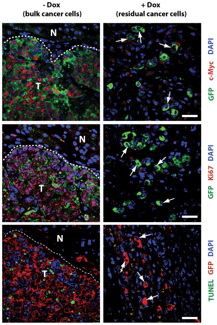
Fig. 2. Dormant cancer cells do not express c-Myc, lack nuclear staining of Ki67, and are TUNEL negative.
Upper panels: Immunofluorescence (IF) staining of c-Myc (red, nuclear) and GFP (green, cytoplasmic) in pancreatic cancers (−Dox, left) and residual cancer cells following tumor regression in response to downregulation of c-Myc (+Dox, right); arrows in the right panel indicate c-Myc-negative nuclei within residual cancer cells; T, tumor; N, adjacent normal tissue (left panels). Middle panels: IF staining of Ki67 (red, nuclear) and GFP (green, cytoplasmic), arrows indicate Ki67-negative nuclei within residual cancer cells. Lower panels: TUNEL labeling (green) of nuclei of apoptotic cells and IF staining against GFP (red, cytoplasmic), arrows indicate TUNEL-negative nuclei within residual cancer cells; bars in all panels represents 25 μm.

