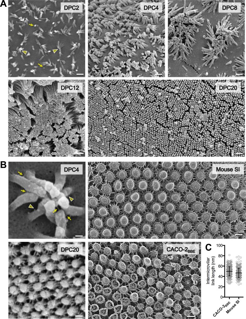
Figure 1. Enterocyte BB microvilli cluster during differentiation and are connected by thread-like links.
(A) SEM of CACO-2BBE cells from a differentiation time series; days post confluency (DPC) are indicated. Yellow arrows point to initial microvillar membrane buds, arrowheads to distal tip contact of longer microvilli. Scale bars, 500 nm. (B) High magnification images of CACO-2BBE cells and native intestinal tissue. Left upper: SEM of a microvillar cluster from 4-DPC CACO-2BBE cell. Yellow arrows point to intact intermicrovillar adhesion links, arrowheads to unpaired or broken links. Left bottom: SEM of a 20-DPC CACO-2BBE monolayer. Right upper: Freeze-etch EM of the BB from mouse small intestinal tissue. Right lower: Freeze-etch EM of the BB from 20-DPC CACO-2BBE cells. Scale bars, 100 nm. (C) Intermicrovillar link lengths from CACO-2BBE and mouse intestinal tissue freeze-etch EM images (mean ± SD). See also Figure S1.

