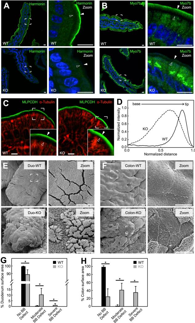
Figure 7. Harmonin KO mice exhibit defects in BB morphology.
(A and B) Confocal images of villi from WT (top) and harmonin KO (bottom) stained for harmonin, Myo7b and DAPI (blue). Boundaries of the BB are denoted with filled arrowheads (microvillar tips) and open arrowheads (terminal web) in the zoomed images (right panels). Scale bars, 10 μm. (C) Confocal images of villi from WT (left) and harmonin KO (right) littermates stained for MLPCDH and α-tubulin. In zoomed images microvillar tips are marked with filled arrowheads and the terminal web is marked with open arrowheads (insets). Scale bars, 5 μm. (D) Line scans of MLPCDH immunofluorescence intensity within the BB of WT and harmonin KO. (E) SEM images of the apical surface of villi from the duodenum (Duod) of WT (top) and harmonin KO (bottom). (F) SEM of the proximal colon apical surface from WT (top) and harmonin KO (bottom) mice. Scale bars, 5 μm. (G and H) Degree of BB defects exhibited in the duodenum (G) and colon (H) of WT and harmonin KO (mean ± SD). No BB defect was defined as an apical surface that possessed well-packed microvilli of uniform length; moderate BB defect as apical microvilli that were disheveled in appearance; severe BB defect as areas that lacked microvilli. Surface area measured: WT SI = 4,001,746 μm2, Harmonin KO SI = 296,867 μm2, WT colon = 511,465 μm2, Harmonin KO colon = 975,557 μm2; n = 4 animals for each genotype. *p value < 0.0001, t test. See also Figure S7.

