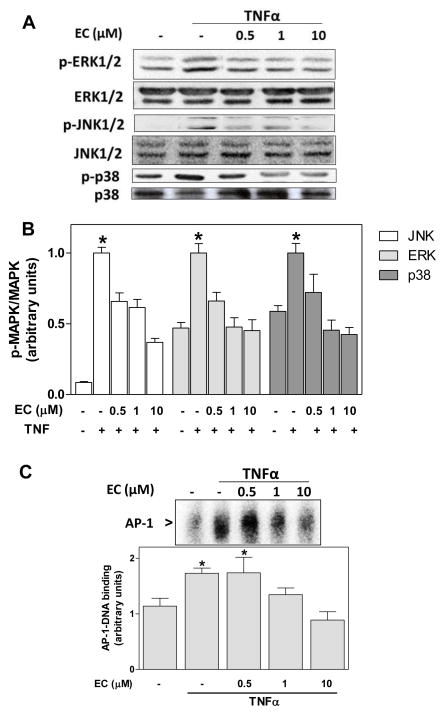
Figure 3. (-)-Epicatechin prevents TNFα-induced MAPK and AP-1 activation in 3T3-L1 adipocytes.
3T3-L1 adipocytes were incubated without or with (-)-epicatechin (EC) (0.5-10 μM) for 4 h, and subsequently in the absence or presence of 20 ng/ml TNFα for further 15 min (MAPKs) or 2 h (AP-1): A- Representative images and B- quantification of Western blots for phosphorylated and non-phosphorylated JNK1/2, ERK1/2, and p38 levels in total cell extracts; C- representative image and quantification of AP-1-DNA binding in nuclear fractions as determined by EMSA. B-C, bands were quantified and results were referred to untreated cell values (Arbitrary unit=1). For Western blots (B) results were expressed as the ratio phosphorylated/non phosphorylated protein levels. Data represent means ± SEM of three to four independent experiments. *Significantly different compared to all other groups (p < 0.05, One way ANOVA test).

