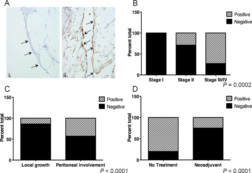
Figure 1. Peritoneal VCAM-1 expression among women with ovarian cancer.
(A) Representative histology of biopsies stained for VCAM-1 expression. Arrows indicate mesothelium. i, example of negative staining; ii, brown staining of mesothelium indicates positive reactivity. (B) Incidence of women with positive VCAM-1 staining of the mesothelium segregated by tumor stage. Omentum or peritoneal biopsies were stained for VCAM-1 using IHC. Specimens were scored positive if >50% of the mesothelial cells showed 3+ reactivity (stippled bars) or negative if <50% of the cells showed reactivity (solid grey). Data represent percent of total patients analyzed (P = 0.0002, Chi-squared test). Stage I, n=12, Stage II, n=14, Stage III/IV, n=22. (C) Percentage of specimens with VCAM-1 positivity among Stage II patients separated based on presence (peritoneal involvement, n=7) or absence (local growth, n=7) of secondary tumor implants within the pelvis (P < 0.0001, Fisher’s Exact test). (D) Percentage of VCAM-1 staining specimens from a set of matched Stage III patients who received upfront surgery (no treatment, n=18) or neoadjuvant chemotherapy (NACT, n=13) (P < 0.0001, Fisher’s Exact test).

