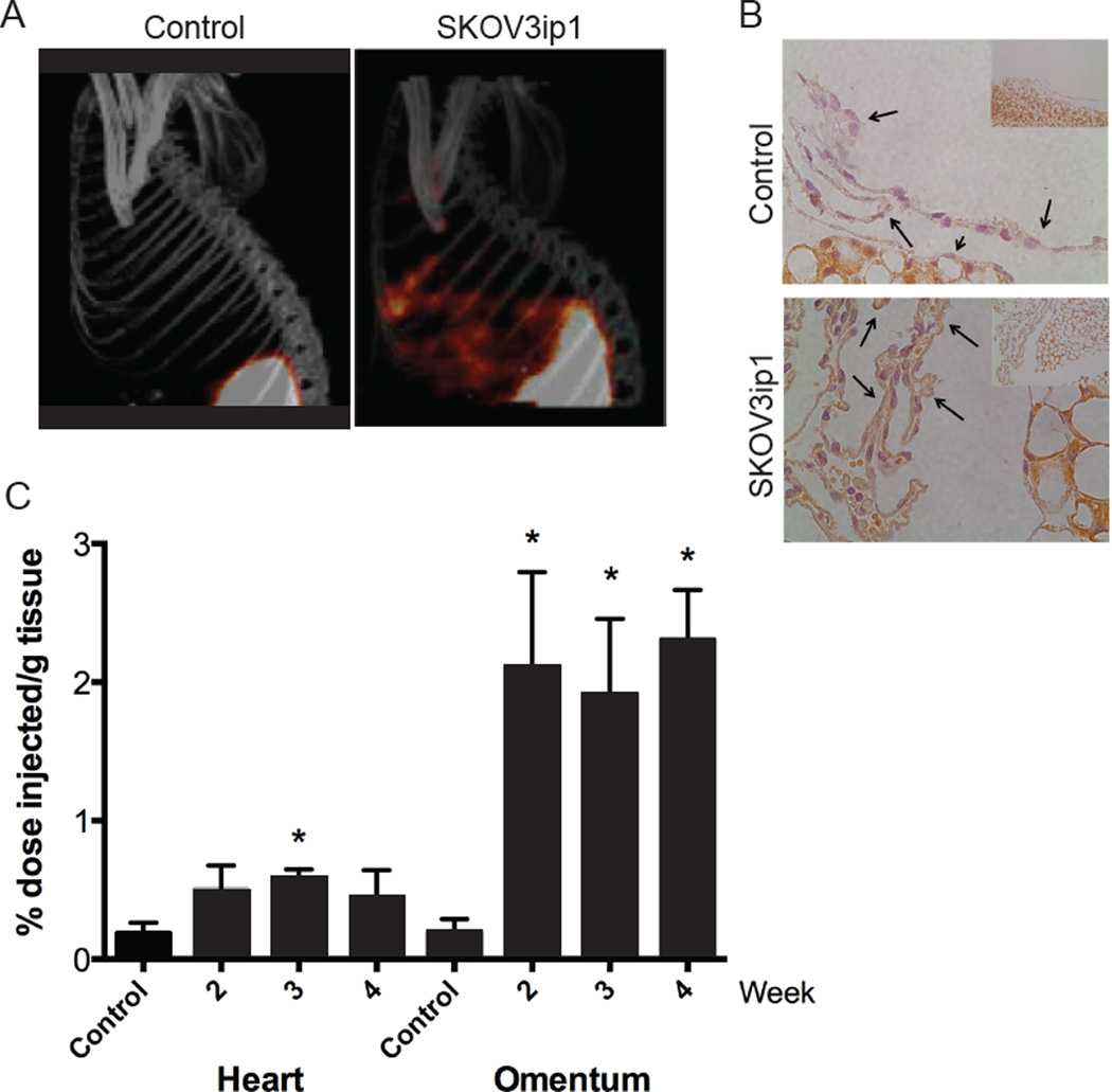
Figure 2. Detection of mesothelial VCAM-1 expression in a mouse model of ovarian cancer peritoneal metastasis.
Athymic nude mice received an intraperitoneal (IP) injection of SKOV3ip1 cells or saline (control). (A) SPECT/CT imaging of 111In-tVCAM-4 peptide 4 hours after IP injection into saline control injected (left panel) or tumor-bearing (right panel) animals. Maximum intensity projection (MIP) images shown. (B) Correlative Histology. Following radioactive decay, the omentums were stained for VCAM-1 using IHC (60X images with 20X insert). Arrows indicate the mesothelium in each case. Top panel – saline injected control mouse; bottom panel – mouse with SKOV3ip1 tumor cells injected. (C) Kinetics of VCAM-1-In111 peptide distribution in the heart or omentum of athymic nude mice 2, 3 and 4 weeks after IP injection of SKOV3ip1 cells, n=3. Control animals were evaluated 2 weeks after PBS injection. Data represent mean ± standard deviation. p<0.05, 1-way ANOVA for heart or omentum samples; *p<0.05, Bonferroni’s Multiple Comparison Test, relative to PBS-injected control for each organ.

