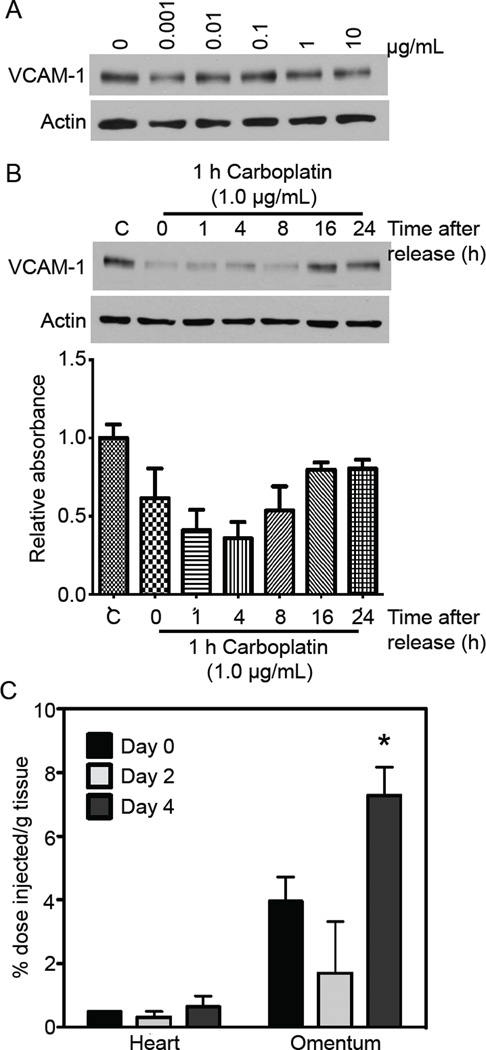
Figure 3. Carboplatin transiently diminishes mesothelial VCAM-1 expression.
(A) LP9 mesothelial cells were treated with the indicated concentrations of carboplatin for 24 hours prior to lysis and immunoblotting for VCAM-1 and actin (loading control). (B) LP9 cells were either untreated or exposed to 1 μg/ml carboplatin for 1 hr prior to release into fresh media. Lysates were blotted for VCAM-1 and actin. Following background subtraction, signal intensities for the VCAM-1 bands were normalized to actin and are expressed relative to the untreated control (C). (C) Nude mice harboring SKOV3ip1 tumors were imaged using SPECT/CT and the 111In-tVCAM-4 peptide 2 weeks post-tumor initiation to obtain baseline distribution values (0). Two days later (2), mice were imaged 8 hours after receiving an IP injection of 25 mg/kg carboplatin and 2 days later; 111In-tVCAM-4 peptide biodistribution presented as mean ± standard deviation.

