
Fig. 1.
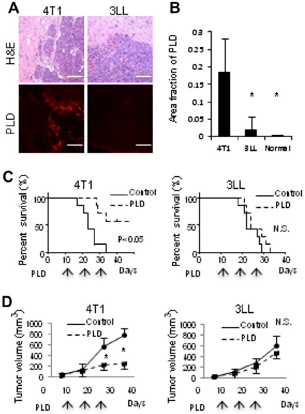
PLD accumulation and therapeutic efficacy. (A) Hematoxylin and eosin (H&E) staining and fluorescence imaging of accumulated PLD in 4T1 and 3LL tumors grown in brain. Bar indicates 100 μm. (B) Quantification of PLD accumulation in tumors. * Indicates p < 0.05 versus 4T1. The therapeutic efficacy of PLD was assessed by analyzing the survival of mice bearing tumors in the brain (C), or by measuring subcutaneous tumor growth (D). The mice were treated three times with PLD or PBS (control) at the times indicated by arrows. N.S. indicates no significant difference in the survival or tumor size. * Indicates p < 0.05 versus control.