
Fig. 4.
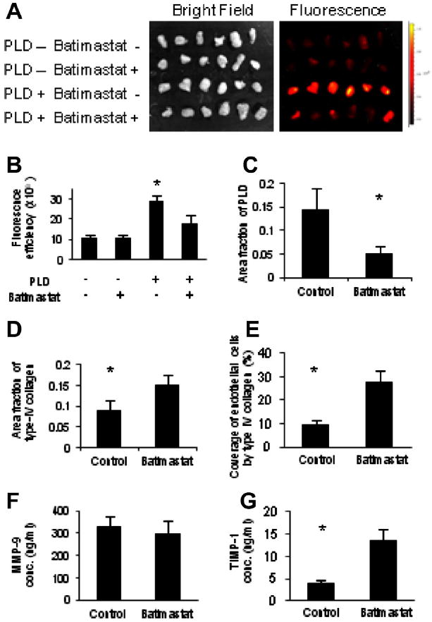
Reduction of vascular permeability to PLD by MMP inhibitor. Batimastat or PBS was injected i.p. once a day for 3 days into the mice bearing 4T1 tumor, prior to i.v. injection of PLD. (A) Accumulation of PLD to the tumors was imaged ex vivo using an IVIS apparatus. (B) Accumulation of PLD to the tumors was abrogated by pre-treatment of mice with Batimastat. * Indicates p < 0.05 versus the other treatments. Confocal microscopic imaging of the tumors and image quantification of PLD accumulation. (C) The amount of type IV collagen. (D) And the coverage of endothelial cells by basement membrane (E). (F) Measurement of serum protein levels by ELISA yielded no significant differences in MMP-9 concentration (conc.). (G) However, a significant increase in TIMP-1 concentration was observed. * Indicates p < 0.05 versus batimastat.