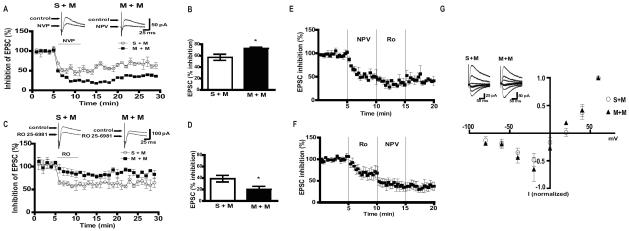
Figure 3.
NMDAR currents are altered in mice that display context-dependent sensitization to morphine. A) (Upper panel) Representative traces and (lower panel) time course of % inhibition of NMDAR EPSCs by NVP in CA1 pyramidal cells from paired S+M and paired M+M mice. B) Summary of % inhibition of EPSCs indicating that neurons recorded in the slices from M+M mice are more sensitive to NR2A subunit antagonist NVP (n=5/group, p<0.05). C) (Upper panel) Representative traces and (lower panel) time course of % inhibition of NMDAR EPSCs by RO 25-6921. D) Summary of % inhibition of EPSCs indicating that neurons from paired M+M mice are less sensitive to RO 25-6921 compared to S+M (n=5/group, p<0.05). E) Average time course of NMDA-EPSCs during the application of NVP followed by RO 25-6981(n=4/group). F) Average time course of NMDA-EPSCs during the application of RO 25-6981 followed by NVP (n=4/group). G) I–V curves and representative traces of NMDAR EPSCs from S+M and M+M mice (n=4–5/group). Data were analyzed using unpaired t-test.

