
Figure 3.
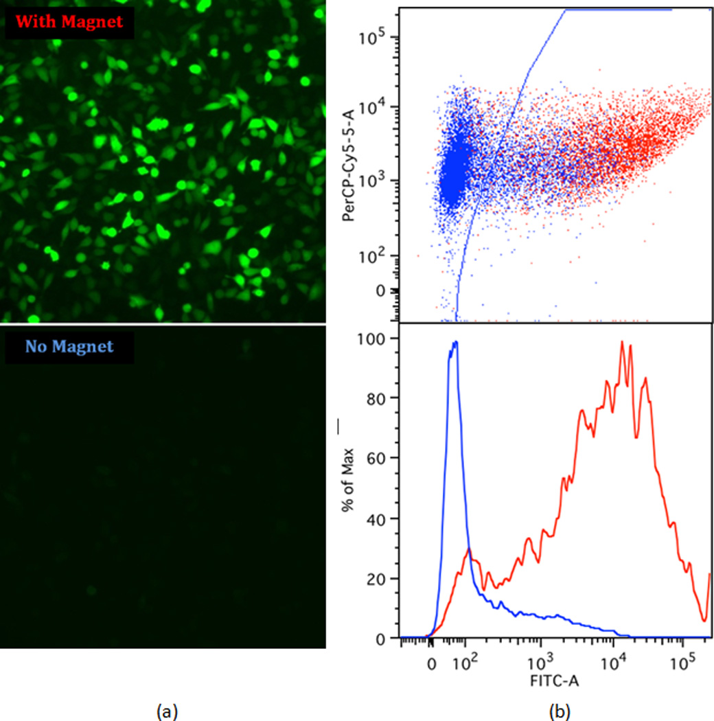
Comparison of delivery efficiency with and without a magnet: (a) fluorescence microscopy images of cells after the transfection of plasmid DNA encoding green fluorescent protein (GFP) with and without the magnetic field; (b) fluorescence-activated cell sorting (FACS) analysis. Based on analysis of non-treated cells, cells to the right of the gate (blue line) were considered GFP-positive, while cells to the left of the gate were fluorescing at levels indistinguishable from background.