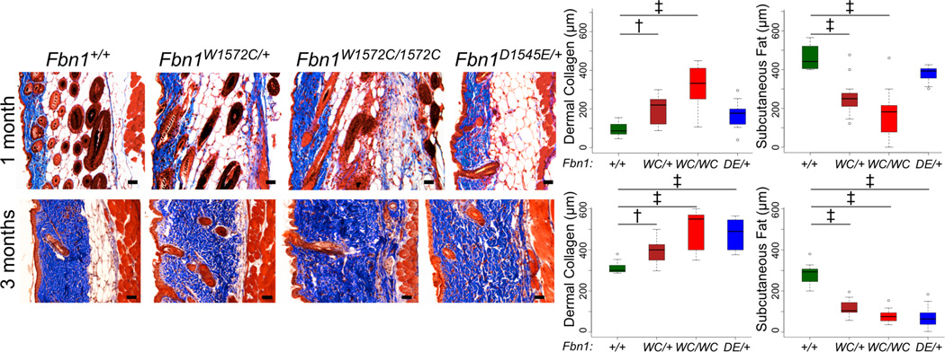
Figure 1. SSS mouse models show skin fibrosis.
Masson’s trichrome staining of back skin sections from male mice (genotypes indicated) at 1 month (top panels) and 3 months (bottom panels) of age demonstrates progressive loss of subcutaneous fat and an expanded zone of dense dermal collagen in mutant animals. Quantification of the thickness of the zones of dermal collagen and subcutaneous fat in wild-type and mutant mice at 1 (top panels) and 3 (bottom panels) months of age is shown. Similar findings were observed in mutant female mice (Figure S2A,B). 1 month males: n = 9 (+/+), 10 (WC/+), 10 (WC/WC), 9 (DE/+); 3 month males: n = 13 (+/+), 9 (WC/+), 9 (WC/WC), 9 (DE/+). Scale bars, 50 µm. * p<0.05, ** p<0.01, † p<0.001, ‡ p<0.0001. DE = D1545E. WC = W1572C.

