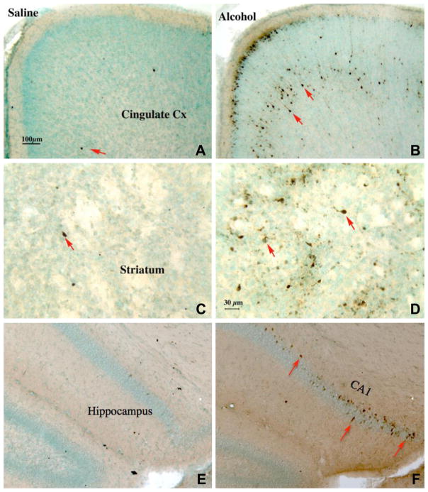
Fig. 1.
Alcohol-induced apoptosis indicated by immunocytochemistry of cleaved-caspase-3, shown in photomicrographs of saline-treated (left panels) and alcohol-treated (right panels) mice for cingulate cortex (top panels), striatum (middle panels), and hippocampal formation (bottom panels). Alcohol was administered to B6 mice on PD 7 (2.5 g/kg twice a day, 2 h apart, s.c.) and the pups were perfused for immunocytochemistry 6 h after the second alcohol injection. In saline-treated mice, very few cleaved-caspase-3 positive neurons were seen (A, C, E; arrows). In contrast, many caspase-3-positive neurons were evident in the cingulate cortex (B) [as well as parietal, temporal and retrosplenial cortices (not shown)], striatum (D), and hippocampal formation CA1/2 [as well as subiculum (not shown)] (F) of mice treated with alcohol. Scale bars: A, B, E, F = 100 μm; C, D = 30 μm.

