
FIGURE 4.
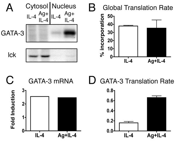
TCR signaling increases the rate of GATA-3 translation. Activated, undifferentiated T cells were restimulated with IL-4 or with both IL-4 and OVA peptide presented by ProAd-B7 cells (Ag+IL-4); hIL-2 was added to both stimulation conditions. A, 48 hours after restimulation T cells were pulsed with 35S met/cys for 10 minutes. Nuclear and cytosolic lysates were immmunoprecipitated for GATA-3 and lck and analyzed on SDS gels. One experiment, representative of 3, is shown. B, The overall translational efficiency was determined by measuring the percent incorporation of 35S met/cys into ethanol-precipitable proteins from the pulse-labeled lysates. Data are depicted as mean + SEM, n=3. C, At the time of the pulse label, mRNA was isolated from a portion of the T cells and measured by real time PCR. The fold induction of GATA-3 mRNA, relative to lck, is shown for the experiment in AD, The GATA-3 translation rate was calculated as the amount of detectable radioactivity from the pulse period divided by the amount of mRNA at the time of the pulse label, both normalized to lck. Data are depicted as mean + SD, p<0.0001 for IL-4 vs Ag+IL-4 by unpaired t test, n=3.