
FIGURE 6.
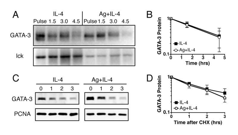
TCR signaling does not increase GATA-3 protein stability. Activated, undifferentiated T cells were restimulated with IL-4 or with both IL-4 and OVA peptide presented by ProAd-B7 cells (Ag+IL-4); hIL-2 was added to both stimulation conditions. A and B, 48 hours after restimulation T cells were pulsed with 35S met/cys for 10 minutes, then chased with unlabeled media for various times. Nuclear and cytosolic lysates were immunoprecipitated for GATA-3 and lck and analyzed on SDS gels. The cell number was adjusted so that the IL-4 and Ag plus IL-4 samples have similar starting levels of GATA-3 protein, to account for the Ag-driven increase in GATA-3 translation. A, One gel, representative of 3, is shown. B, The quantitation of GATA-3 protein level during the pulse chase experiments is depicted as mean +/- SD, n=3. For some experiments the pulse time was increased to 60 minutes. C and D, 100 μg/mL cycloheximide (CHX) was added to T cells and protein levels of GATA-3 and PCNA were measured by western blot of nuclear lysates. C, One western blot, representative of 3, is shown. D, The quantitation of GATA-3 protein level after addition of CHX is depicted as mean +/- SEM, n=3.