
FIG. 1.
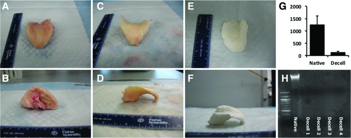
Gross morphologic view of native (A, B), decellularized (C, D), and lyophilized (E, F) tissues. The three-dimensional structure is mostly retained. PicoGreen assay (G) and agarose gel electrophoresis (H) confirm decellularization. Color images available online at www.liebertpub.com/tea