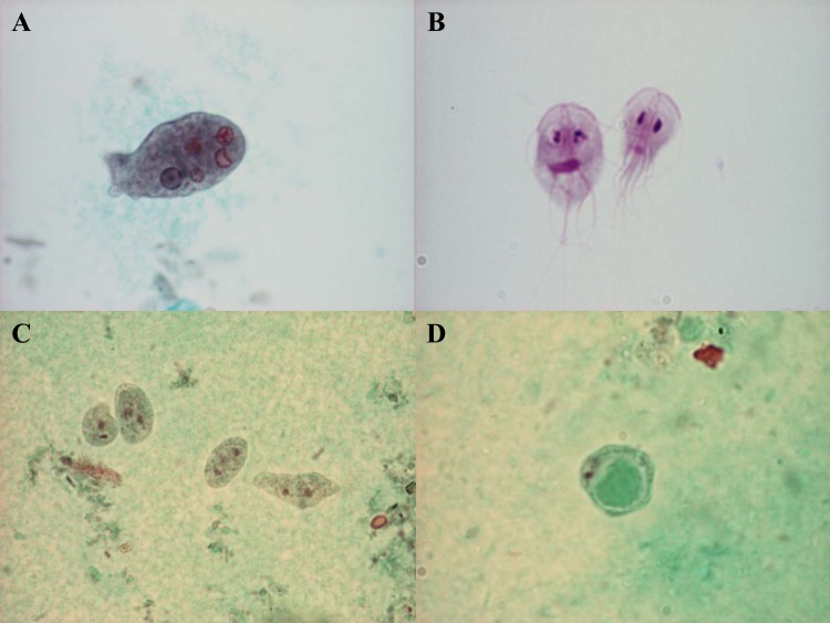
FIG 2.
Trichrome staining of an Entamoeba histolytica trophozoite with ingested red blood cells (A), Dientamoeba fragilis trophozoites (C), and the Blastocystis vacuolar stage (D). (B) Giemsa staining of Giardia lamblia trophozoites. (Parasite images courtesy of Marianne Lebbad; reprinted with permission.)

