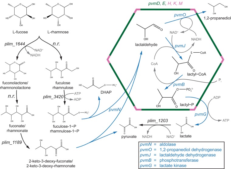
FIG 6.
Proposed model of l-fucose metabolism in P. limnophilus. Two potential pathways to reach lactaldehyde are shown. Enzymes encoded by the pvm gene cluster are shown in blue, and shell proteins are shown in green (BMC-H) or pink (BMC-P). Cofactors are depicted in gray. Enzymes that potentially catalyze reactions not directly involved in the BMC chemistry are shown in black. “n.r.” stands for no result, meaning that there was no significant BLAST hit using characterized enzymes as a query. DHAP, dihydroxyacetone phosphate.

