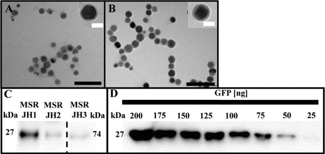
FIG 4.

(A and B) Transmission electron micrographs of purified magnetosomes from M. gryphiswaldense ΔC JH3 (A) and wt (B), showing no effect on MM or magnetite crystals. Black scale bar, 200 nm; white scale bar, 40 nm. (C) Quantitative Western blot of (Mag-)EGFP in the MM, isolated from M. gryphiswaldense strains expressing different chromosomal mamC–Mag-egfp fusions from PmamDC45 (JH1) and Ptet (JH2) and mamC–Mag-egfp–egfp fusions (JH3) from PmamDC45. (Mag-)EGFP was detected by rabbit anti-GFP IgG as the primary antibody and goat anti-rabbit IgG horseradish peroxidase antibodies as secondary antibodies. The sample containing the Mag-EGFP–EGFP fusion was diluted 10× for quantitative Western blot analysis. Band sizes are as follows: GFP, 27 kDa; 2× GFP, 74 kDa. (D) Recombinant GFP was used as a standard.
