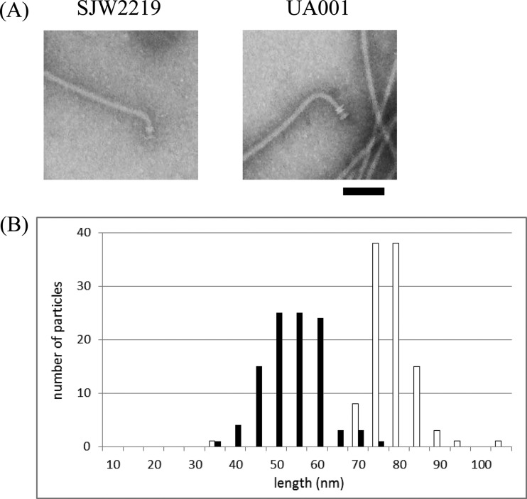
FIG 1.
(A) Electron micrographs of flagellar basal structures from strains UA001 (right) and SJW2219 (left). Intact flagella were isolated from cells grown in TY medium at 30°C as previously described (10). The hook length of UA001 is significantly greater than that of SJW2219. Samples were negatively stained with 2% phosphotungstic acid (pH 7). Bar, 100 nm. (B) Hook length distribution of intact flagella isolated from strains UA001 (empty bars) and SJW2219 (filled bars) grown at 30°C. The number of particles counted was 105 for UA001 and 101 for SJW2219.

