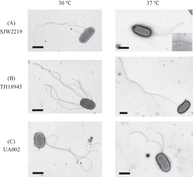
FIG 2.

Electron micrographs of SJW2219 (A), TH18945 (B), and UA002 (C) cells grown in TY medium at either 30 or 37°C. In an enlarged image of the SJW2219 flagellar basal structures in the membrane, both intact and immature basal structures are visible (A, inset). Cells were negatively stained with 1% phosphotungstic acid (pH 7). Bars, 1 μm.
