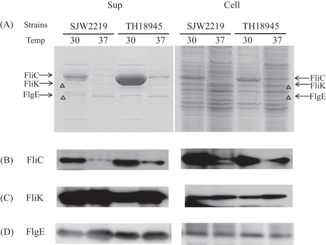
FIG 3.

(A) SDS-PAGE pattern of proteins secreted into the culture medium (Sup) by SJW2219 and TH18945 cells (Cell). Cells were grown in TY medium either at 30 or 37°C. Secreted proteins were recovered from the cultures by pelleting the cells and precipitating the supernatant with TCA (Sup). Whole cells were collected from the pellets (Cell). The gels were stained with CBB. The acrylamide concentration of the gel was 12.5%. The proteins secreted by SJW2219 cells grown at a temperature (Temp) 30°C are shown in lane 1, while those of cells incubated at 37°C are shown in lane 2. Secreted proteins from TH18945 cells grown at 30°C are shown in lane 3, and those from the same cells grown at 37°C are in lane 4. Proteins from the pellets of SJW2219 cells grown at 30°C are in lane 5, and those from the same cells grown at 37°C are in lane 6. Proteins from pellets of TH18945 cells grown at 30°C are in lane 7, and those from the same cells grown at 37°C are in lane 8. (B) Western blot analysis of the supernatant and cellular fractions of SJW2219 and TH18945 cells with anti-FliC polyclonal antibodies at a 1:20,000 dilution. Cells were grown at 30 and 37°C to an OD600 of 1.0. Samples in lanes are the same order as those in panel A. (C) Detection of intracellular and extracellular FliK with polyclonal anti-FliK antibodies (1:20,000 dilution). Samples in lanes are in the same order as those in panel A. (D) Detection of intracellular and extracellular FlgE with polyclonal anti-FlgE antibodies (1:20,000 dilution). Samples in lanes are in the same order as those in panel A.
