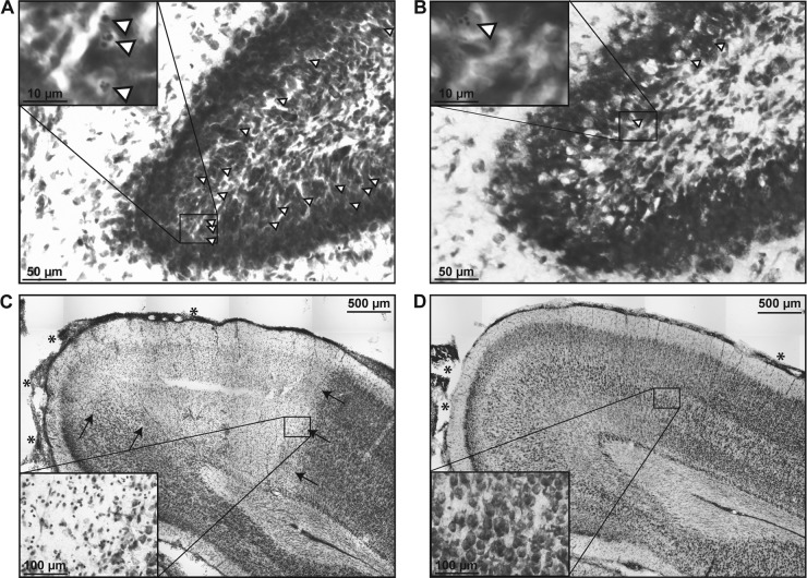
FIG 3.
Representative images (Nissl staining) of cryosections prepared 42 h after the induction of PM in infant rats. (A) Apoptotic neurons (selected cells are marked with arrowheads; inset) were found throughout the hippocampal DG of animals treated with the vehicle and ceftriaxone, whereas apoptotic cells were found only sporadically in animals treated with Ro 32-7315 and ceftriaxone (B). (C) A sharply demarcated necrotic zone in the cortex (arrows) with meningeal leukocyte infiltration (asterisks) is visible in an animal treated with the vehicle and ceftriaxone, as opposed to the intact cortex with slight meningeal leukocyte infiltration (asterisks) in an animal treated with Ro 32-7315 (D).

