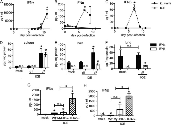
FIG 1.
Reduced IFN-γ production and elevated type I IFNs in a mouse model of fatal ehrlichiosis. Mice were infected with E. muris or IOE, and levels of IFN-γ and type I IFNs (α and β) were measured by ELISA at different times postinfection as indicated. Serum IFN-γ (A), IFN-α (B), and IFN-β (C) concentrations in WT C57BL/6 mice infected with E. muris (closed circles) or IOE (open circles) are shown. IFN-α (closed bars) and IFN-β (open bars) in the spleens (D), livers (E), and lungs (F) of IOE-infected WT mice at day 7 postinfection are shown. (G) IFN-α concentration in the sera of IOE-infected WT (closed bars), MyD88−/− (open bars), and TLR2−/− (striped bars) mice at day 7 postinfection is shown. (H) IFN-β in the sera of IOE-infected WT, MyD88−/−, and TLR2−/− mice at day 7 postinfection is shown. Asterisks indicate a significant difference compared to the counterpart mock controls (P < 0.05). “n.s.” indicates no significant difference (P > 0.05) and “n.d.” indicates not detectable. Number symbols (#) indicate a significant difference between groups as indicated (P < 0.05). At least three animals were used per group.

