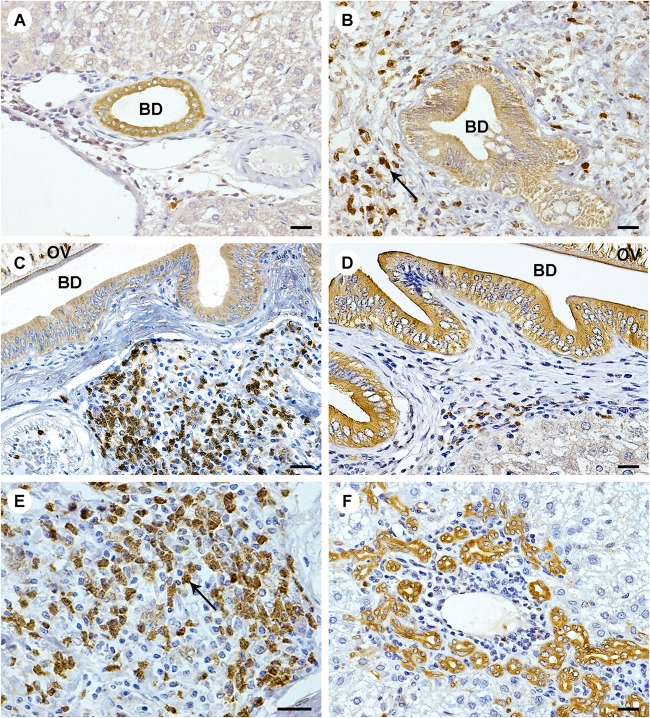
FIG 5.
Immunohistochemistry of ANXA1 expression in livers of hamsters infected with O. viverrini. Immunohistochemistry was performed in normal hamster liver at 5 months (A) and in O. viverrini-infected livers at 1 month (B), 3 months (C), and 5 months (D). A single representative image is shown for each time point (5 liver samples per time point). ANXA1 was expressed in the cytoplasm of inflammatory cells (arrow [B and E]) and proliferating bile duct epithelial cells (F). The magnification is ×400, except in panel E, where it is ×600 (scale bar = 200 μm). BD, bile duct; OV, Opisthorchis viverrini.

