
FIG 2.
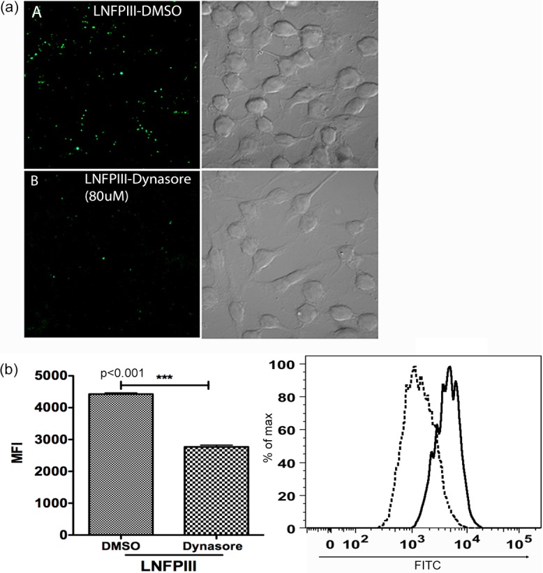
Endocytosis of LNFPIII-NGC is dynamin mediated. (a) Raw 264.7 cells were pretreated with 80 μM dynasore (panel B) or an equal volume of carrier molecule DMSO (panel A) for 40 min at 37°C and then incubated with 50 μg/ml LNFPIII-NGC for 30 min at 37°C. Cells were harvested, fixed, and stained for internalized LNFPIII-NGC (green) using E.5 antibody followed by incubation with Alexa Fluor 488-labeled secondary antibody. Cells were visualized, and images were taken with a Zeiss LSM 510 META inverted confocal microscope under a 60× objective. (b) Raw 264.7 cells were pretreated with 40 μM dynasore for 40 min and were then incubated with 50 μg/ml of LNFPIII-NGC at 4°C for 10 min. Endocytosis of LNFPIII was induced for 20 min at 37°C. Cells were harvested, fixed, and stained for internalized LNFPIII as described before. Cells were acquired on a BD LSRII flow cytometer and analyzed by FlowJo software. Mean fluorescence intensity (MFI) for FITC was measured for live cells gated as SSC- and FSC-positive populations. Data are from three independent experiments performed. Treated and nontreated samples were in triplicate in each independent experiment. Black solid line, LNFPIII (DMSO); black dotted line, LNFPIII (dynasore). Statistical analysis was performed using Student's t test (***, P < 0.001).