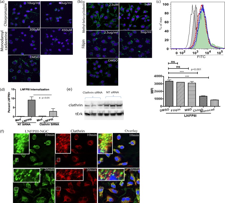
FIG 3.
Endocytosis of LNFPIII-NGC is clathrin mediated. (a and b) Raw 264.7 cells were pretreated with clathrin inhibitors chlorpromazine (10 and 40 μg/ml) and monodansylcadaverine (300 and 450 μM) or caveolus inhibitors methyl-β-cyclodextrin (2.5 and 5 μM) and filipin (2.5 and 5 μg/ml) for 40 min at 37°C. The cells were further incubated with 50 μg/ml of LNFPIII-NGC. Endocytosis was induced for 30 min at 37°C, and then the cells were fixed and stained for internalized LNFPIII-NGC as described in Materials and Methods. Nuclei were stained with Hoechst dye (blue). (c) Under similar conditions as for panels a and b, FACS analysis of LNFPIII-NGC uptake by cells treated with clathrin and caveolus inhibitors was performed. Different histograms indicate LNFPIII-NGC + DMSO in solid blue line, LNFPIII + MBD (methyl-beta cyclodextrin) as a filled green histogram, LNFPIII + filipin in red dotted line, LNFPIII + monodansyl cadaverine as a filled gray histogram, and LFPIII + Chlrph (chlorpromazine) in a thin black line. FITC mean fluorescence intensity (MFI) was measured for live cells gated as SSC- and FSC-positive populations. Cells were acquired on a BD LSRII flow cytometer and analyzed by FlowJo software (Tree Star Inc.). Statistical calculation was performed using Student's t test analysis (***, P < 0.001; **, P < 0.01). Inhibitor-treated and -nontreated samples were in triplicate, and data represent 3 independent experiments performed. (d) siRNA-mediated knockdown of endogenous expression of clathrin reduced LNFPIII-NGC uptake by APCs. RAW 264.7 cells were transfected with clathrin-specific siRNA or nontarget siRNA for 36 h. Transfected cells were incubated with LNFPIII-NGC at 37°C for 30 min and immunolabeled with E.5, followed by incubation with Alexa Fluor 488-conjugated secondary antibody. Cells were then acquired and analyzed via flow cytometry as described before. The data are representative of 3 independent experiment with n = 4. Statistical analysis was performed using Student's t test (*, P < 0.05). (e) Western blot showing downregulated expression of clathrin protein. Thirty-six hours posttransfection with clathrin siRNA, cell lysates were run on SDS-PAGE, and the corresponding Western blots were probed with anticlathrin antibody. Total Erk was used as a loading control. (f) LNFPIII-NGC colocalizes with endogenous clathrin. Endocytosis of LNFPIII-NGC was induced in RAW 264.7 macrophages for 10 and 20 min at 37°C. Cells were fixed and double stained for LNFPIII-NGC and clathrin protein as described in Materials and Methods. Images were captured using a Nikon Eclipse Ti A1R confocal microscope system. Endocytosed LNFPIII-NGC is in green, clathrin is in red, and an overlay image of colocalized LNFPIII-NGC and clathrin is in yellow. The zoomed-in image of part of a cell is kept as an inset for better representation.

