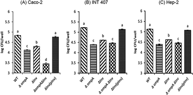
FIG 2.

C. sakazakii ATCC 29544 invasion of Caco-2 (A), INT-407 (B) and Hep-2 (C) cells. Confluent cell monolayers were infected with bacteria at an MOI of 40 and incubated for 1.5 h, followed by a gentamicin protection assay. The cells were treated with 0.25% Triton X-100 to release the bacteria, and intracellular bacteria were enumerated. The data represent the average log CFU per well ± the standard deviation (SD) from at least three independent experiments performed in duplicate. Values of P of <0.05 were considered significantly different, and bars with the same lowercase letter are not significantly different from one another.
