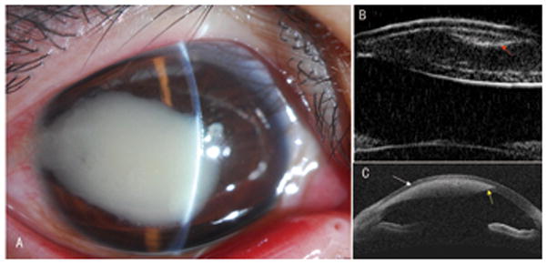
Figure 2. AS-OCT and UBM for imaging intra-stromal corneal foreign bodies (a bamboo spur).

A: A 20-year-old man had a dense corneal leukoma secondary to an infiltrative reaction of an intra-stromal corneal foreign body in his right eye.
B: A UBM image showed both boundaries of the cornea and a clear curved foreign body with hyper-reflection (red arrow) beneath the anterior face of the corneal lesion. The corneal lesion presented with two surfaces in the cornea.
C: An OCT image showed the anterior face of an oval shadow with focal strong hyper-reflection inside in the corneal stroma (white arrow) and just a small part of the posterior face at the periphery (yellow arrow).
