
FIG 3.
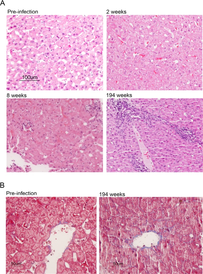
Liver histopathology in chimpanzee 4x0193. (A) Hematoxylin and eosin-stained sections of tissue collected prior to and following infection with H77S.2 virus. Mild hepatocellular swelling with minimal impingement of hepatic sinuses and disruption of cords is evident at 2 weeks postinoculation of virus. Following the appearance of viremia, 8 weeks after inoculation, there was moderate hepatocellular swelling with increased impingement of sinuses. Minimal lymphocyte infiltration was evident in portal regions. By 194 weeks, the hepatic cords had become disorganized and the sinusoidal spaces markedly narrowed. Periportal mononuclear cell infiltrates were increased, and there was diffuse moderate hepatocellular swelling with increased amounts of pale, glassy cytoplasm, mild anisocytosis, occasional binucleate hepatocytes, and rare single-cell necrosis. (B) Masson trichrome stains of preinfection (left) and week 194 (right) liver tissue showing evidence of fibrosis (collagen, blue) with septae lining hepatic sinusoids in tissue collected at 194 weeks.