
FIG 4.
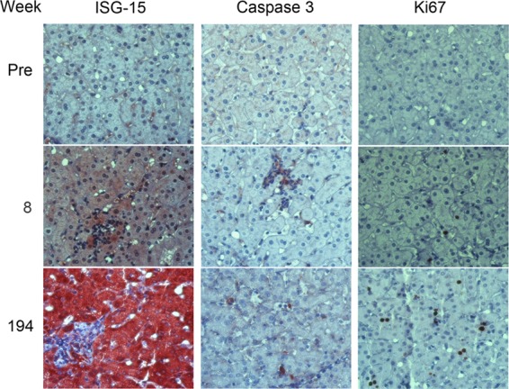
Immunohistochemical staining of 4x0193 liver tissue for markers of innate immune response (ISG-15), apoptosis (caspase 3), and cellular proliferation (nuclear Ki67 expression). A marked increase in hepatocellular ISG-15 expression is evident between preinfection liver and tissue collected at 8 and especially 194 weeks. Caspase 3 expression is also increased, especially in association with collections of lymphoid cells. Multiple, single, and paired Ki-67-positive nuclei are evident at 194 weeks, consistent with increased hepatocellular proliferation.