
FIG 3.
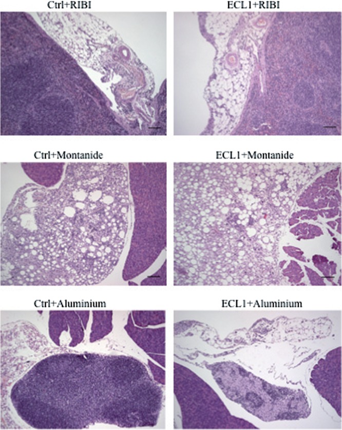
Representative histopathology analysis performed on perisplenic adipose tissues (top row), peripancreatic adipose tissues (middle row), and lymph node tissues (bottom row) from mice receiving ECL1 antigen in complete formulation or as the sole immunization adjuvant. All the images show moderate to severe chronic inflammation of the adipose tissue. RIBI plus ECL1, protocol O; Montanide plus ECL1, protocol M; aluminum plus ECL1, protocol N.