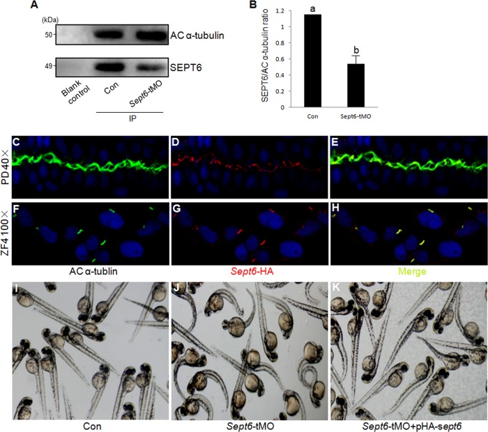
FIG 10.
SEPT6 associates with acetylated α-tubulin in cilia. (A) The endogenous SEPT6 coimmunoprecipitates with acetylated (AC) α-tubulin. Briefly, lysates from embryos were immunoprecipitated with resin beads coated with anti-acetylated α-tubulin. The levels of acetylated α-tubulin (upper panel) and SEPT6 (lower panel) in the precipitates were analyzed by Western blotting using antibodies against each protein. Lane 1, blank control sample; lane 2, control embryos; lane 3, sept6-tMO morphants. (B) Quantification of SEPT6 that coimmunoprecipitated with acetylated α-tubulin (α-tubulin also serving as the normalization standard) from the control embryos and sept6-tMO morphants. The letters a and b indicate statistically significant differences (P < 0.001, calculated by using Student's t test). (C to E) Localization of HA-SEPT6 in relation to the acetylated microtubules in the cilia of the pronephric ducts in embryos injected with pHA-sept6, visualized by double staining with antibodies against acetylated α-tubulin (C) and the HA tag (D). Acetylated α-tubulin and SEPT6 localizations are merged in panel E. (F to H) Colocalization of HA-SEPT6 with acetylated α-tubulin in the cilia of pHA-sept6-transfected ZF4 cells. Acetylated α-tubulin (F) and HA-SEPT6 (G) showed colocalization as indicated in the merged image (H). (I to K) Coinjection of the HA-sept6 plasmid at 15 ng/μl effectively rescued the global defects of embryos that were caused by sept6-tMO.

