FIG 6.
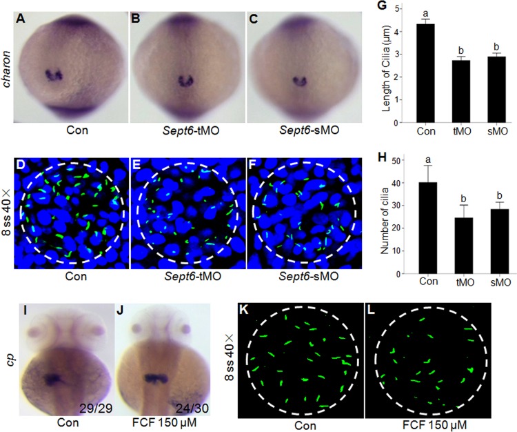
Knockdown of sept6 causes defects in KV ciliogenesis. (A to C) Representative images of charon expression in KV in control embryos (A), sept6-tMO morphants (B), and sMO morphants (C). (D to F) Cilia in the KV were visualized by immunofluorescence of acetylated α-tubulin in the control embryos (D), sept6-tMO morphants (E), and sMO morphants (F). (G and H) Bar charts represent the quantification and statistical analyses of cilia length (G) and numbers (H) at the 8-ss stage. The letters a and b represent statistically significant differences (P < 0.001, calculated by using Student's t test). (I and J) FCF treatment caused defects in LR patterning of the liver, as demonstrated by WISH analysis of the liver-specific marker cp at 50 hpf in the control (I) and FCF-treated (J) embryos. (K and L) Immunofluorescence of acetylated α-tubulin in the control (K) and FCF-treated (L) embryos. Nuclear DNA was stained with 4′,6-diamidino-2-phenylindole, and the KV is circled (D to F, K, and L).

