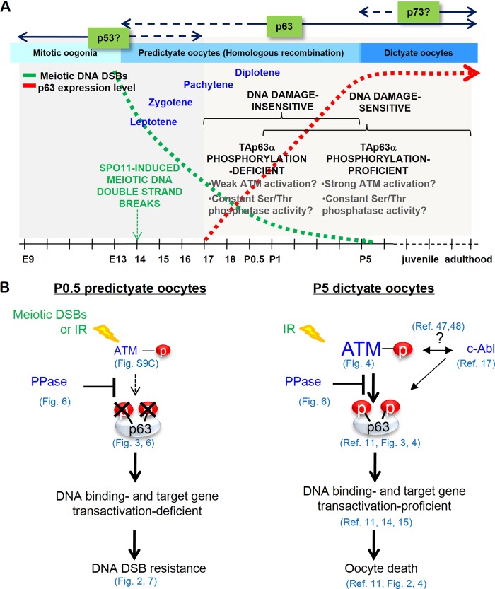
FIG 7.
Summary and model of TAp63α expression and phosphorylation regulation and DNA damage sensitivity changes during oocyte development. (A) Timeline outlining the increase in TAp63α phosphorylation potential and DNA damage sensitivity in oocytes in the context of prophase I events during female germ line development. Meiotic DNA DSB levels decline (green dotted line) through homologous recombination-mediated DNA repair as TAp63α expression levels rise in oocytes (red dotted line), starting from E18.5 and peaking at P5. Note that the “DNA damage-sensitive” and “DNA damage-insensitive” refer to only immature primordial follicle oocytes. (B) Model showing how relative activities of the p63 phosphorylation ATM kinase and TAp63α phosphorylation downregulating Ser/Thr protein phosphatase (PPase) in P0.5 versus P5 oocytes could determine the final level of TAp63α phosphorylation induction and oocyte survival or death upon meiotic or IR-induced DNA DSB formation.

