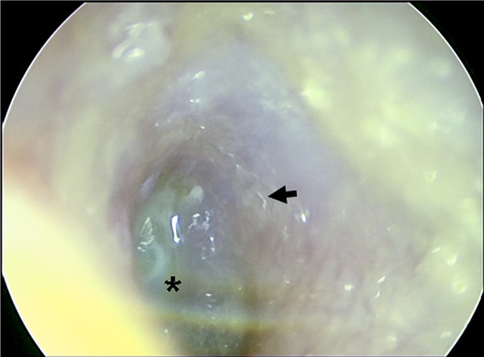
FIG 1.

Otomicrosocopy of the external auditory canal. An endoscopic view into the left outer ear canal onto the tympanic membrane with the central umbo (*) and the atypical cone of light in the upper half is shown. Larvae can be discerned as undulatory light reflections (arrow).
