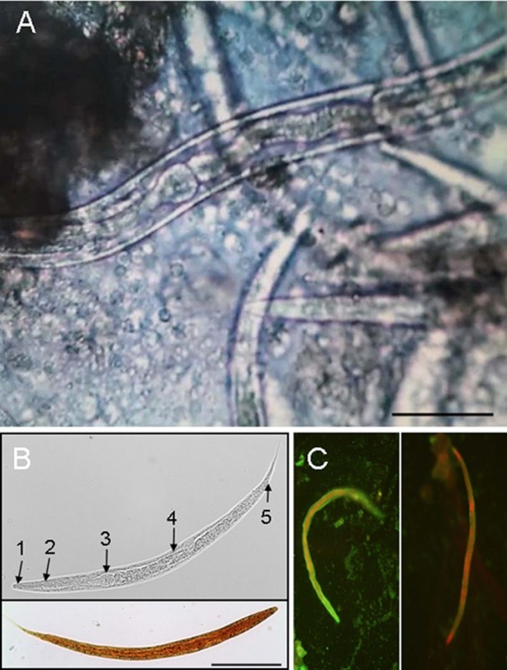
FIG 2.

Microscopic visualization of Rhabditis larvae in ear canal lavage fluid. (A) Unstained microscopy of the ear canal lavage fluid. Bar, 50 μm. (B) Unstained (upper panel) and iodine-stained (lower panel) male larvae. Bar, 100 μm. Upper panel: 1, stoma/buccal cavity; 2, esophagus; 3, esophageal bulb; 4, intestine; 5, anus. (C) Indirect immunofluorescence staining of larvae in the lavage fluid using the patient's serum (left panel [diluted 1:10]) or serum obtained from a healthy blood donor (right panel [diluted 1:10]).
