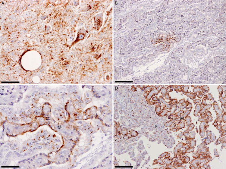
FIG 1.
(A) Dorsal motor nucleus of the vagus nerve from ewe PG1657/05 showing widespread PrPSc deposits. (B) Placentome from fetus PG1658/05 at the 4th month of pregnancy showing restricted PrPSc deposits. (C) Greater magnification of the PrPSc-positive area shown in panel B to demonstrate that both maternal and fetal opposing units are affected and to illustrate the different PrPSc deposition patterns between maternal and fetal units. These different PrPSc deposition patterns most likely reflect the different physiological roles of maternal and fetal units, as both units were of the same PrP genotype. (D) For comparison, a placentome collected near term demonstrates the widespread PrPSc dissemination at the end of the 5th month of gestation. All sections were labeled with the monoclonal antibody R145. The maternal unit is denoted by the letter M, the fetal unit by the letter F. The scale bars in panels A and C represent 50 μm, in panels B and D, 200 μm.

