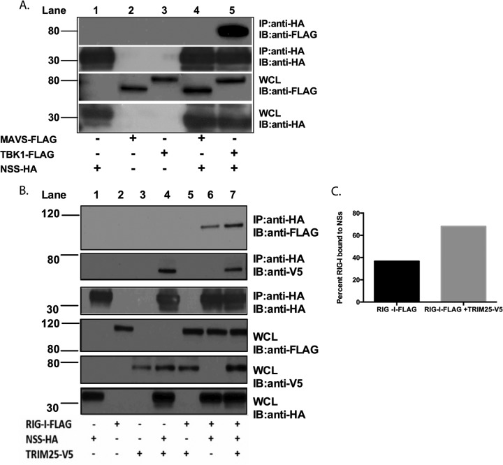
FIG 3.
SFTSV NSs interacts with TBK1, RIG-I, and TRIM25. (A) HEK293T cells were transfected with the indicated expression plasmids, and at 24 h after transfection, cell lysates were collected and subjected to immunoprecipitation (IP) by using anti-HA antibodies. Immunoprecipitates were analyzed by immunoblotting (IB) using anti-FLAG and anti-HA antibodies. Whole-cell lysates (WCL) were immunoblotted by using anti-FLAG and anti-HA antibodies. (B) HEK293T cells were transfected with the indicated expression plasmids, and at 24 h after transfection, cell lysates were collected and subjected to immunoprecipitation by using anti-HA antibodies. Immunoprecipitates were analyzed by immunoblotting using anti-FLAG, anti-V5, and anti-HA antibodies. Whole-cell lysates were immunoblotted by using anti-FLAG, anti-V5, and anti-HA antibodies. Numbers to the left of the gels in panels A and B are molecular masses (in kilodaltons). (C) Densitometry analysis of immunoprecipitated RIG-I for panel B. The band signal intensity of the immunoprecipitated RIG-I protein was normalized to the signal intensity of the RIG-I protein expressed in the whole-cell lysate. Signal intensities were obtained by using Image Studio Lite software.

