FIG 4.
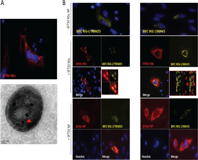
SFTSV NSs targets the RIG-I/TRIM25 complex to cytoplasmic structures. (A) HeLa cells were transfected with a plasmid carrying SFTSV NSs fused to mCherry, and at 24 h after transfection, cells were fixed and analyzed by confocal microscopy (top). In another set of experiments, the SFTSV NSs-induced structures were purified from cells stably expressing SFTSV NSs-mCherry using serial centrifugation and OptiPrep density gradient centrifugation and subjected to immunogold labeling and electron microscopy analyses (bottom). Red arrowhead, SFTSV NSs protein staining. (B) HeLa cells were transfected with BiFC RIG-I/TRIM25 (left) or BiFC RIG-I/MAVS (right) construct pairs along with an empty plasmid or a plasmid carrying SFTSV NSs-HA. Transfected cells were incubated for 24 h at 37°C and then incubated at 30°C for 2 h to promote fluorophore maturation. Cells were fixed and analyzed by confocal microscopy. Rabbit anti-HA and Alexa Fluor 633 were used for detection of SFTSV NSs. Cell nuclei were visualized by using Hoechst dye.

