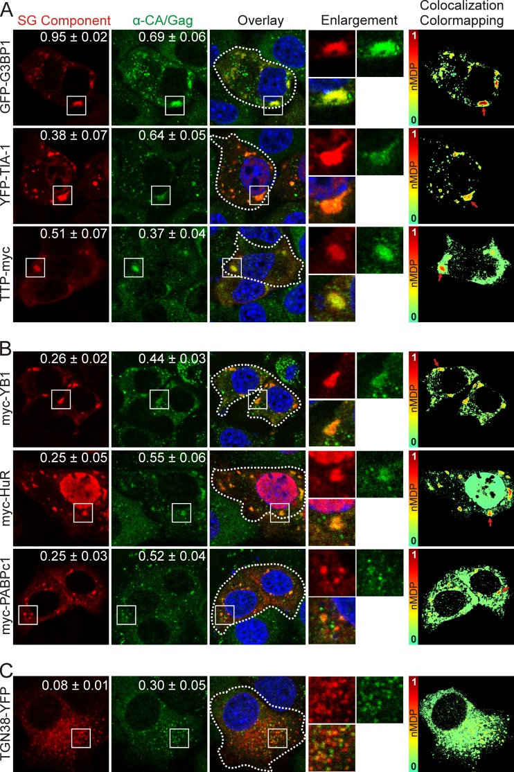
FIG 2.
Expression of stress granule-associated proteins relocalizes MMTV Gag to stress granules. Confocal microscopy images show MMTV Gag localization with core stress granule proteins that nucleate assembly (A), proteins that localize to stress granules but have not been reported to nucleate stress granule assembly (B), and a control protein that localizes to the trans-Golgi network and does not induce stress granule formation (C). White boxes indicate the areas shown in the enlargements. To the right, colocalization color maps were generated of the cells outlined by white dashed lines using the colocalization color map plugin (48) for ImageJ (http://imagej.nih.gov/ij/). Red arrows point to discrete regions of high colocalization on the color-mapping images. SG, stress granule.

