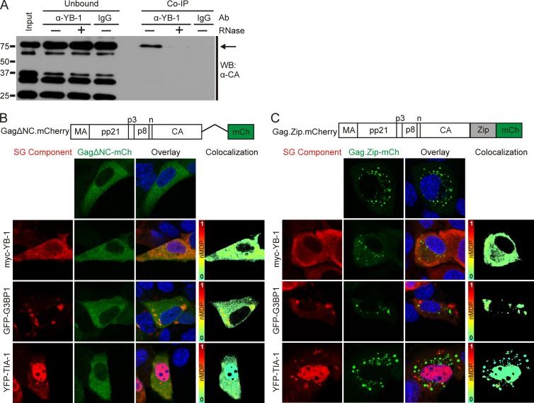
FIG 5.
RNA-dependent interaction of Gag with stress granule proteins. (A) Endogenous YB-1 was immunoprecipitated from MMTV(C3H)-infected NMuMG whole-cell lysates using 5 μg of YB-1 antibody or an irrelevant IgG antibody as a control. Prior to the immunoprecipitation, samples were treated with (+) or without (−) RNase A. Gag (indicated by the arrow) was detected by Western blotting (WB) using anti-CA monoclonal antibody (Ab). (B) Schematic diagram showing the NC deletion mutant Gag.ΔNC-mCherry with images of cells expressing the protein (false colored green in the images) expressed alone or coexpressed with myc–YB-1, GFP-G3BP1, or YFP–TIA-1 in uninfected NMuMG cells. Colocalization color maps were generated as described in the legend of Fig. 4 and are shown to the right. (C) Schematic representation of the Gag.Zip-mCherry construct, in which the NC domain of Gag was replaced with the CREB1 leucine zipper domain, with images of cells expressing the construct (false colored green) alone or with myc–YB-1, GFP-G3BP1, or YFP–TIA-1 in NMuMG cells. Colocalization color maps are shown to the right. mCH, mCherry.

