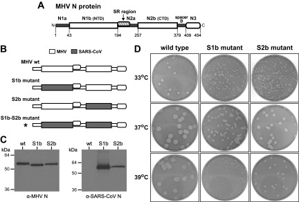
FIG 1.

Construction of MHV mutants with chimeric N proteins containing RNA-binding domains from the SARS-CoV N protein. (A) Model of the MHV N protein showing domains as defined previously (16, 17). The RNA-binding domains are N1b (or NTD) and N2b (or CTD). Other segments of the molecule include the amino terminus N1a; the central spacer N2a, which harbors the SR region that interacts with nsp3; and the M protein-binding domain N3, which is linked to the molecule via spacer B. Numbering indicates amino acid residues. (B) Schematic of wild-type and mutant N proteins. Shading represents segments of SARS-CoV N sequence substituted within the MHV N molecule; the asterisk denotes a lethal substitution. (C) Western blots of equal amounts of immunopurified wild-type, S1b mutant, and S2b mutant virions probed with monoclonal anti-MHV N antibody J.3.3, which recognizes an epitope in domain N3 (31) (left) or with polyclonal anti-SARS-CoV N antibody (right). (D) Plaques of the S1b and S2b mutants at 33, 37, or 39°C compared with those of isogenic wild-type virus. Plaque titrations were carried out on L2 cells; monolayers were stained with neutral red at 49 h postinfection and were photographed 17 h later.
