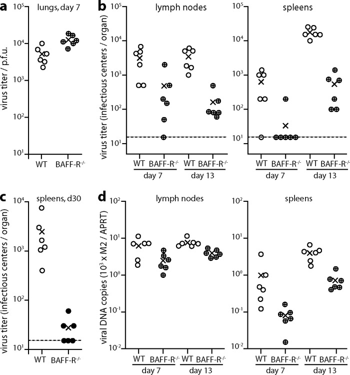
FIG 1.
Impaired colonization of BAFF-R−/− mice by i.n. MuHV-4. (a) C57BL/6J (WT) or BAFF-R−/− mice were infected i.n. with MuHV-4 (104 PFU). Seven days later, titers of infectious virus in lungs were detemined by plaque assay. Circles show individual mice; crosses show means. BAFF-R−/− lung titers were significantly higher than those of WT mice (P < 0.01). (b) Mice were infected as for panel a, and virus loads in mediastinal lymph nodes and spleens were determined 7 and 13 days later by infectious center assay. Circles show individual mice; crosses show means. The dashed lines indicate the lower limit of assay sensitivity. At day 13, WT titers were significantly higher than those of BAFF-R−/− mice in both lymph nodes (P < 0.001) and spleens (P < 0.0001). (c) Mice were infected as for panel a, and virus loads in spleens were determined 30 days later by infectious center assay. Circles show individual mice; crosses show means. The dashed line indicates the lower limit of assay sensitivity. WT titers remained significantly higher than those of BAFF-R−/− mice (P < 0.05). (d) The organs of mice for panel b were assayed for viral DNA by Q-PCR. Viral genome loads (M2) are normalized by the cellular genome load of each sample (APRT). At day 13, viral DNA loads were significantly higher in WT spleens and lymph nodes than in those of BAFF-R−/− mice (P < 0.003).

