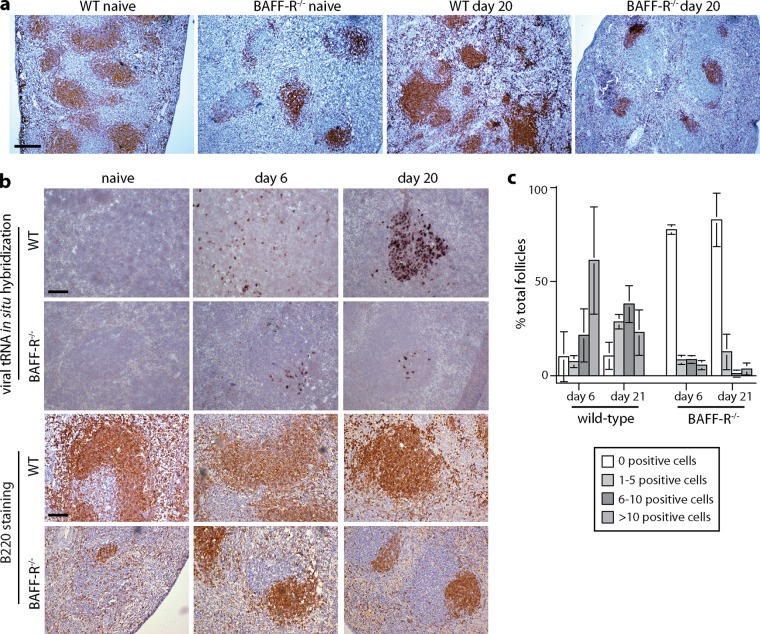
FIG 4.
Defective GC formation in MuHV-4-infected BAFF-R−/− mice. (a) Spleen sections from C57BL/6J (WT) and BAFF-R−/− mice were stained for B220 (brown) either before (naive) or 20 days after i.p. MuHV-4 (105 PFU) and counterstained with Mayer's hemalum. The scale bar shows 500 μm. The number of B220 follicles per section was significantly lower in BAFF-R−/− (mean ± standard deviation [SD], 4.7 ± 1.4; n = 6) than in WT (14.2 ± 1.5) spleens, both before and after infection (P < 10−6). (b) We infected C57BL/6J (WT) and BAFF-R−/− mice i.p. with MuHV-4 (105 PFU) and then detected viral tRNA/miRNA transcripts by in situ hybridization (dark staining). Examples of positive follicles are shown. B220 staining of the same spleens (brown) relates the extent of tRNA/miRNA staining to the extent of the white pulp occupied by B cells. Scale bars show 100 μm. (c) tRNA/miRNA+ cells were counted for spleen sections from 3 mice per group, and follicles were scored as uninfected or infected at a low (1 to 5 cells per section), medium (5 to 10 cells per section), or high (>10 cells per section) level. Bars show means ± SD, counting 20 to 30 follicles per mouse. At both day 6 and day 20, WT spleens contained significantly fewer tRNA/miRNA+ follicles than BAFF-R−/− spleens (P < 0.0001 by 2-tailed Fisher's exact test), and among the positive follicles, the number of tRNA/miRNA+ cells was significantly higher in WT spleens (P < 0.01 by Student's unpaired 2-tailed t test).

