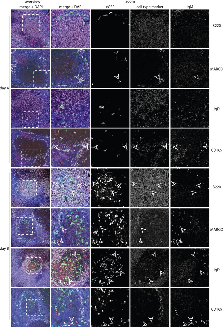
FIG 5.
Colonization of C57BL/6J splenic follicles by eGFP+ MuHV-4. We infected C57BL/6J mice i.p. with MuHV-4 expressing eGFP from an intergenic EF1α promoter (105 PFU) and 4 or 8 days later identified infection by staining splenic sections for eGFP (green) plus IgM (red; mainly marginal-zone B cells) and, as a further cell type marker, either B220 (mainly follicular B cells), MARCO (marginal-zone macrophages), IgD (mainly follicular B cells), or CD169 (marginal-zone metallophilic macrophages) (white). Nuclei were counter-stained with DAPI (blue). The boxed regions of the left-hand overviews (scale bars show 100 μm) are shown at higher power in the right-hand zoomed images, with either merged or individual channels. Arrows show examples of eGFP+ cells.

