
FIG 7.
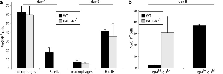
Quantitation of splenic colonization by eGFP+ MuHV-4. (a) We infected C57BL/6J (WT) or BAFF-R−/− mice i.p. with eGFP+ MuHV-4 as for Fig. 5 and 6 and after 4 or 8 days typed eGFP+ cells as macrophages (CD169+ or MARCO+) or B cells (IgM+ or IgD+). Each bar shows the mean ± SD for 3 sections from each of 3 mice. Macrophage infection was not significantly different between the mouse groups, but WT mice showed significantly more B cell infection at day 4 (P < 0.001). (b) EGFP+ B cells at day 8 were further subdivided into IgMhi IgDlo (predominantly marginal-zone) and IgMlo IgDhi (predominantly follicular) populations. The eGFP+ B cells of WT mice were significantly higher in IgMlo IgDhi and significantly lower in IgMhi IgDlo than those of BAFF-R−/− mice (P < 0.001).