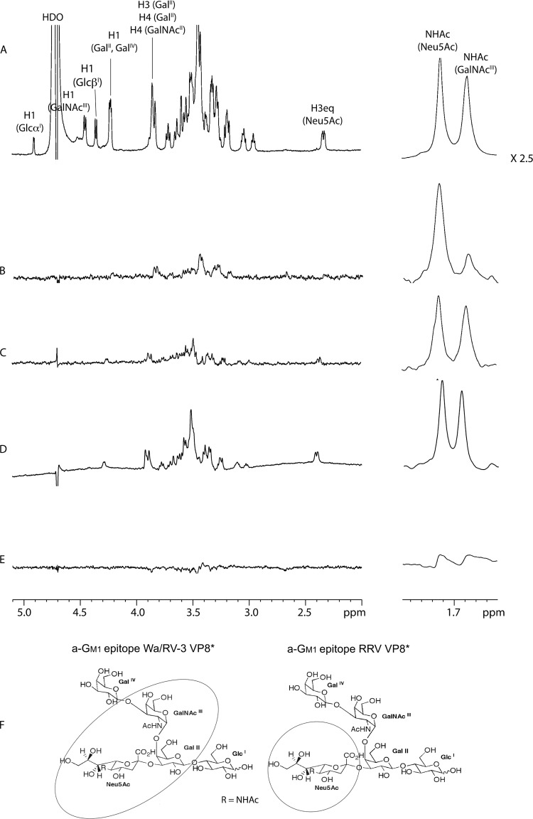
FIG 4.
NMR spectra of a-GM1. (A) Control 1H NMR spectrum of a-GM1. STD NMR spectra of a-GM1 in the presence of RRV GST-VP8* (B), Wa GST-VP8* (C), RV-3 VP8* (D), and with GST alone (E) are shown. In all panels, the signals of the two N-acetamido groups' methyl protons at approximately 1.70 ppm are depicted separately on the right at a uniform magnification of ×2.5. All spectra were acquired in deuterated phosphate buffer at 600 MHz and 280 K. (F) Structure of a-GM1 with the binding epitopes of VP8* encircled. The Wa and RV-3 VP8* epitopes include both the Neu5Ac and GalNAc residues (left), whereas the RRV VP8* epitope encompasses only the Neu5Ac moiety (right).

