
FIG 6.
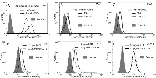
Flow-cytometric analysis of MA104 cell surface GM1 availability and effect of CTB on cell binding by VP8* of Wa, RV-3, and CRW-8. (A) Trypsin-EDTA treatment reduced cell surface GM1 availability. Histograms indicate the GM1 surface exposure, determined by incubation with FITC-CTB, on cells that were placed into suspension by trypsin-EDTA or scraping and rested for 30 min. Each cell preparation was reacted with unconjugated FITC for the controls, which each showed similar histograms. (B and C) Cell binding by recombinant VP8* of Wa (B) and RV-3 (C). Wa and RV-3 GST-VP8* proteins were tested for binding at concentrations (μg/ml) of 5, 50, 150, and 300 to cells placed into suspension using trypsin-EDTA treatment. Effect of cellular treatment with CTB or inactivated (inact.) CTB at 1 μg/ml on cell binding by VP8* of Wa (D), RV-3 (E), and CRW-8 (F). Wa, RV-3, or CRW-8 GST-VP8* protein was added at 300 μg/ml to cells placed into suspension with trypsin-EDTA. (D to F) The histogram of each VP8* bound to untreated cells was indistinguishable from that of the corresponding VP8* bound to cells treated with inactivated CTB.